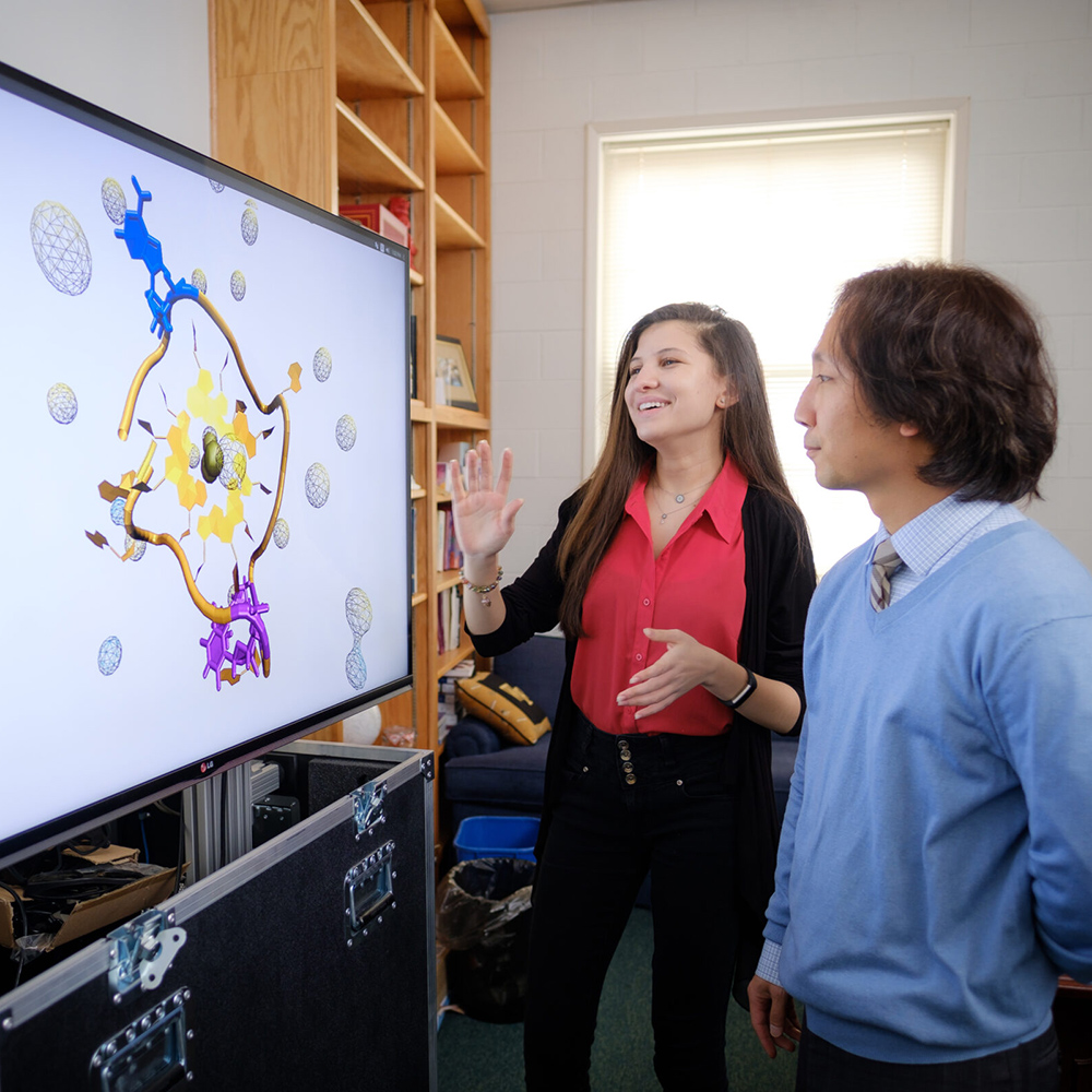
Biophysics is an interdisciplinary science that applies the principles and methods of physics to understand biological systems. It involves studying the physical aspects of molecules, cells, and organisms, and how these physical properties influence their function and behavior. By integrating methods from physics, biophysicists can explore complex biological phenomena, such as protein dynamics, protein folding, cellular dynamics, and the mechanics of biomolecular interactions. Much of the biophysics at Wake Physics focuses on disease-relevant studies.
SCHOLARSHIP
Faculty working in Biophysics

Daniel B. Kim-Shapiro
Professor of Physics and Harbert Family Distinguished Chair for Excellence in Teaching and Scholarship
Biophysics
Experimental and some computational biophysics, nitric oxide, hemoglobin and other heme proteins, blood flow, and Sickle cell and other cardiovascular diseases
208 Olin Physical Laboratory | Lab: 216 Olin Physical Laboratory

Keith Bonin
Professor of Physics and Richard T. Williams Faculty Chair in Physics
Biophysics
Optics, biophysics, and microscopy
Office: 310 Olin Physical Laboratory | Lab: 200 Olin Physical Laboratory

Sam Cho
Associate Professor and Shively Family Faculty Fellow
Biophysics
Computational biophysics, protein and RNA folding, biomolecular assembly, molecular machines, and GPU-based programming
301B Olin Physical Laboratory | Manchester 228

Martin Guthold
Department Chair, Professor of Physics
Biophysics
Atomic force and fluorescence microscopy of biological samples, and mechanical properties of biological fibers and molecules
Office: 302 Olin Physical Laboratory | Lab: 213 Olin Physical Laboratory
Jed Macosko
Professor of Physics
Biophysics
Mechanics of protein machines
Office: 215 Olin Physical Laboratory | Lab: 213 Olin Physical Laboratory
Fred Salsbury
Physics Graduate Program Director, Professor of Physics
Biophysics
Computational and theoretical biophysics, molecular physics, and drug discovery
301A Olin Physical Laboratory | Lab: 305B Olin Physical Laboratory
Daniel Kim-Shapiro
Daniel Kim-Shapiro leads a laboratory that uses a variety of biophysical and biological techniques to understand blood flow. The lab studies how the important signaling molecule nitric oxide is compromised in various disease states, stored blood, and various cardiovascular disorders and ways to restore nitric oxide in these conditions. The work focuses on basic mechanistic science but also involves translation to the clinic including several clinical trials.
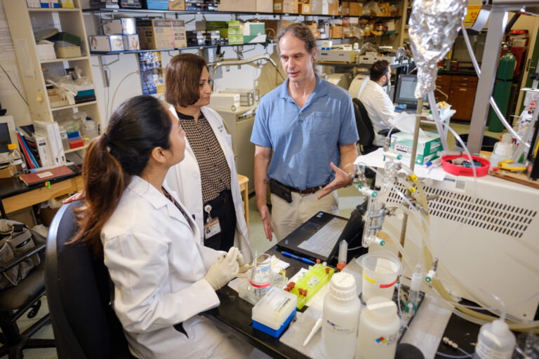

Accepting graduate students
Accepting undergraduate students
Keith Bonin
Keith Bonin’s highly collaborative research has been focused on optics and biophysics with other professors within the Physics Department and the Wake Forest School of Medicine (WFSOM). Currently research includes, among others: studying mechanical properties of cells in 2D, optogenetics studies of the mesolimbic system in the brain, and studies on the motion of double-strand breaks in DNA in cancer cell nuclei.

Not accepting graduate students
Accepting undergraduate students
Sam Cho
Sam Cho’s interdisciplinary research group interests encompass biophysics and computer science. They are interested in the theoretical and computational studies and methods development of biomolecular coarse grained and atomistic molecular dynamics (MD) simulations in collaboration with experimental groups.
Accepting graduate students
Accepting undergraduate students
Martin Guthold
Martin Guthold’s lab’s research interests are in the general areas of biophysics, molecular biology, nanotechnology, microscopy techniques, – especially atomic force microscopy (AFM) and fluorescence microscopy, and next generation sequencing. Current projects include, among others, studies of mechanical and structural properties of fibrin fibers and blood clots and physical properties of cancer cells and normal cells.
Accepting graduate students
Accepting undergraduate students
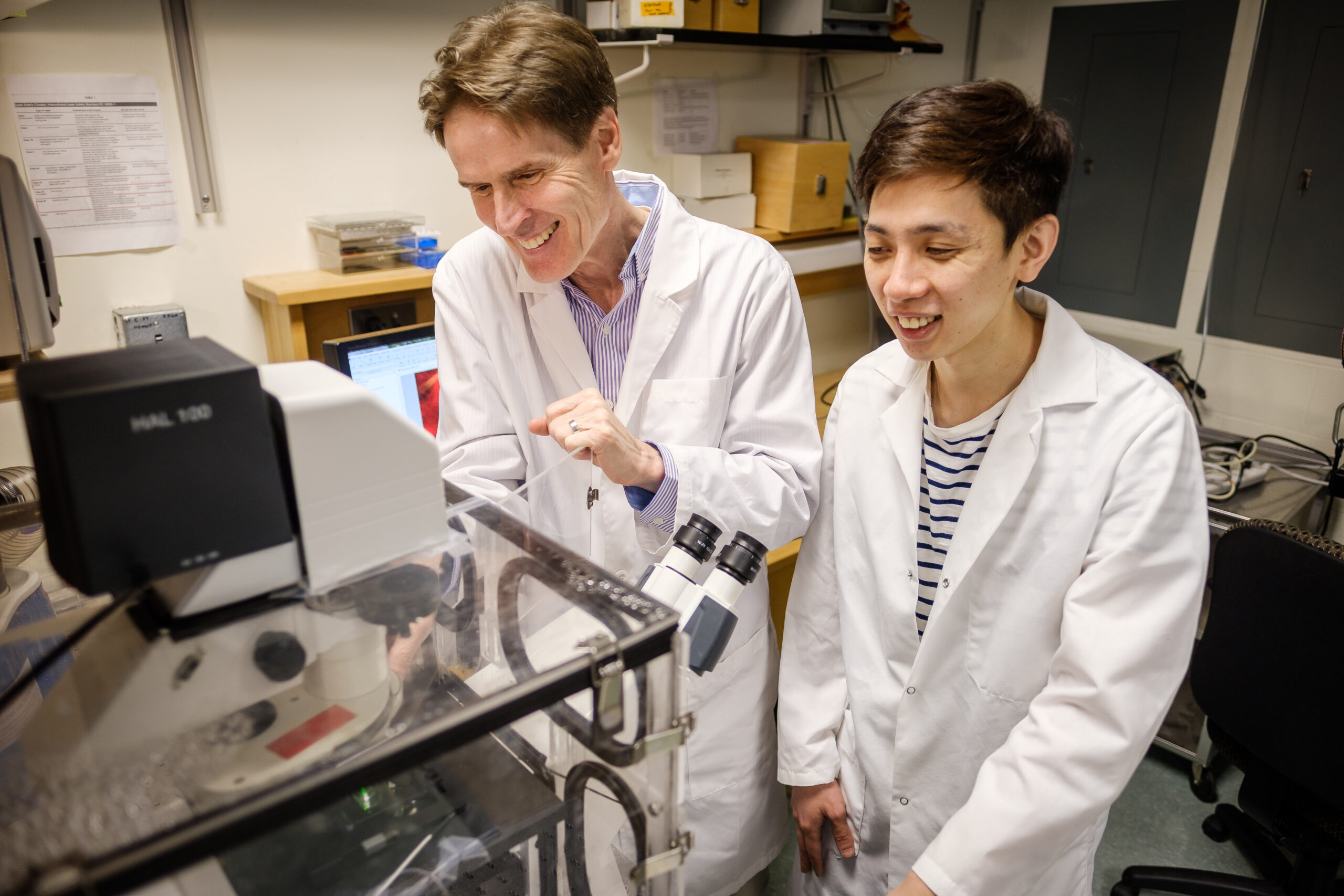
Jed Macosko
Jed Macosko’s group’s long-term goal is the identification of precise mechanical-chemical couplings in molecular machines and the characterization of the overall pathways of their physical motion. They use a varieties of microscopies such as atomic force microscopy (AFM), single molecule fluorescence microscopy and video-enhanced differential interference contrast light microscopy (VE-DIC).
Accepting graduate students
Accepting undergraduate students
Fred Salsbury
Fred Salsbury’s research aims to help in the fight against disease by applying tools from computational physics to molecular-scale problems. Current projects focus on: 1) MD simulations with GPUs 2) drug discovery for chemotherapeutic development 3) development and application of new statistical methods 4) application of machine learning to macromolecular dynamics and drug development. 5) studies of dynamic allostery computationally.
Accepting graduate students
Accepting undergraduate students
Recent Publications in Biophysics
- Nichols, S. L., Hollis, T., Salsbury, F. R., & Esstman, S. M. (2025). K294e change in the rotavirus factory forming protein NSP2 stabilizes a rare C-terminal conformation. Journal of Biomolecular Structure and Dynamics, 1–17. https://doi.org/10.1080/07391102.2025.2563689
- Smith, K. C., Oglietti, R., Moran, S. J., Macosko, J. C., Lyles, D. S., & Holzwarth, G. (2024). Directional change during active diffusion of viral ribonucleoprotein particles through cytoplasm. Biophysical Journal, 123(17), 2869–2876. https://doi.org/10.1016/j.bpj.2024.04.025
- Flake, C., McPherson, H. R., Ariëns, R. A. S., Hudson, N. E., Maultsby, H., Lopez, L., & Guthold, M. (2025). Exploring the functional role of tandem repeats in fibrin(ogen) αc region. Journal of Thrombosis and Haemostasis, 23. https://doi.org/10.1016/s1538-7836(25)00775-5
- Cho, S. S., & Salam, A. (2025). Understanding the role of short- and long-range intermolecular interactions in novel computational drug discovery. Expert Opinion on Drug Discovery, 20(11), 1419–1432. https://doi.org/10.1080/17460441.2025.2555271
- Bonin, K., Prasad, S., Caulkins, W., Holzwarth, G., Baker, S. R., & Vidi, P.-A. (2022). Three-dimensional tracking using a single-spot rotating point spread function created by a multiring spiral phase plate. Journal of Biomedical Optics, 27(12). https://doi.org/10.1117/1.jbo.27.12.126501
- Rochon, E., DeMartino, A., Liu, R., Xu, Q., Kim-Shapiro, D., & Gladwin, M. (2025). In vivo conversion of ferric heme to no-ferroheme has vasodilatory and anti-inflammatory effects. Circulation, 152(Suppl_3). https://doi.org/10.1161/circ.152.suppl_3.4373706